
 العربية
  Azərbaycan
  Kurdish
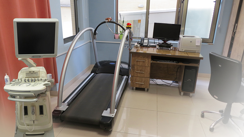
Echocardiography
Echocardiography, also known as a cardiac echo test, is a test in which sound waves are used to create moving images of the heart. Hospital admission is not required for echocardiography. The procedure is painless and is not a surgical operation. Echocardiography is a test used to help the physician diagnose cardiac problems or assess how the heart is functioning. Echocardiography may be necessary in the following circumstances:
- When abnormal heart sounds are present.
- When unexplained chest pain occurs.
- When there is a congenital heart defect.
- After a heart attack.
- In cases of rheumatic fever.
How is Echocardiography Performed?
- You lie on a bed on your side or on your back.
- The physician applies a special gel to a probe and moves it over the chest area.
- Ultrasound waves create images of the heart and its valves.
- X-ray waves are not used in echocardiography.
- Your heart movements are observed on a video screen.
- A video or photograph can be recorded.
- You may also view the video during the test.
- The test usually requires less than 15 to 20 minutes.
- The test is painless and has no side effects.
- Your physician will discuss the results with you.
The cardiac echocardiography service for esteemed clients is located in the cardiac clinic of the Nobar Building (formerly Nasr) of Behboud Specialty and Subspecialty Hospital.
Exercise Stress Test
The exercise stress test of Behboud Hospital is located in the cardiac clinic of the Nobar Building (formerly Nasr) and is operated by experienced experts ready to serve esteemed clients.
In the early stages of coronary artery obstruction in patients with stable angina, there is usually no problem at rest, and symptoms of coronary artery disease often appear during physical activity. Therefore, in order to more accurately and realistically evaluate blood flow to the heart muscle, it is preferable to assess the heart under conditions of activity and stress, so that any region experiencing relative reduction in blood flow (ischemia) due to arterial stenosis (atherosclerosis) can be more clearly identified. With timely treatment of the stenosis, disease progression and subsequent risks can be prevented. When a person experiences activity-related cardiac pain, the resting electrocardiogram may show no changes; in such cases, the exercise test largely resolves this ambiguity. In other words, the exercise test reveals latent changes in the resting electrocardiogram by imposing stress on the heart through physical exercise and activity.
Patient Preparation Before the Exercise Test
You must fast for 3 hours before the test or avoid heavy meals, and you must not have smoked. It is recommended to wear comfortable clothing and shoes, and to perform light warm-up activity before the test, but not strenuous physical activity. (If you have diabetes, fasting for one hour after a light meal is sufficient, and there is no need to change the insulin dose; however, you must inform your physician.) Note that certain cardiac medications that prevent the heart rate from increasing during exercise (such as digoxin, propranolol, metoprolol, atenolol, diltiazem, and others) must be discontinued at a specific interval before the exercise test, with the knowledge and supervision of your treating physician.
Test Procedure
To perform the test, the pulse and blood pressure are first measured and recorded; the patient is connected to ECG electrodes and a baseline electrocardiogram is obtained. The patient is then placed on a treadmill for exercise (running). The treadmill is a moving rubber surface (similar to a conveyor belt) on which the patient stands and walks or runs at the speed of the belt. Every 3 minutes, the incline and speed of the belt increase according to a predefined program. The device starts slowly but gradually increases in speed, requiring the patient to run at the corresponding pace. Gradually, the device tilts upward, creating the sensation of running uphill. The patient’s vital signs and ECG are monitored in different conditions to identify changes in waves and segments during exercise. Based on age and sex, a defined level of exercise and a target maximum heart rate are determined before the test, and running continues until that point is reached. If the heart rate reaches at least 85% of the maximum expected heart rate for the patient’s age and sex during the test, the results can be interpreted. The most reliable result is obtained when the patient reaches 100% of predicted activity without chest pain, severe shortness of breath, or ECG changes, which usually requires about 10 to 12 minutes of exercise. Nevertheless, the test must be discontinued early at any time if the patient feels unable to continue, or if symptoms such as chest pain, severe shortness of breath, dizziness, severe headache, abnormal ECG changes, or a drop in blood pressure occur. For some elderly patients or those with joint or muscular pain who are unable to perform the exercise test, or for those whose baseline ECG changes prevent interpretation of new ECG changes, alternative methods such as cardiac radioisotope scan with pharmacological stress (thallium test) or stress echocardiography should be used instead of exercise. Cardiac scans and stress echocardiography are easier and more accurate than the exercise test but are more expensive.
Applications of the Exercise Test
Overall, the exercise test is a procedure that can detect coronary artery stenoses (greater than 60%) and guide the physician’s decision regarding more invasive procedures (such as angiography). Other applications include evaluating the functional capacity (exercise tolerance) of patients who have suffered a heart attack or have heart failure, in order to determine permissible activity levels.
Limitations of the Exercise Test
In some cases (depending on the affected vessel), the exercise test can produce a false-negative result in up to 25% of cases (a negative report despite coronary obstruction). Conversely, the exercise test may produce false-positive results in healthy individuals. The exercise test also cannot detect mild atherosclerotic stenoses; although these atherosclerotic plaques have lower clinical significance, they may sometimes rupture and cause a heart attack.
Contraindications of the Exercise Test
Active ischemia, recent heart attack, or certain ECG changes that prevent interpretation of results are among the contraindications. Additionally, when the patient is unable to exercise for any reason or exercise is deemed harmful, certain medications are injected to induce stress, followed by cardiac imaging methods (stress echocardiography or nuclear scan).
Electrocardiography
Electrocardiography, or recording the electrocardiogram, is a method for evaluating the electrical activity of the heart by recording the path of an electrical impulse from its origin in the sinoatrial node, which causes the heart to contract. An electrocardiogram (ECG) is a graph that displays the electrical activity of each heartbeat as well as the rhythm of successive heartbeats.
The electrocardiograph is a device that receives the electrical signal of cardiac contraction during one heartbeat cycle from the surface of the skin. Its electrodes are placed at specific locations on the skin, and the electrical signal is displayed as a curve on paper or on a monitor. It is referred to by the abbreviation ECG (Electrocardiogram).
The electrocardiography unit is located in the emergency department and the cardiac clinic, and is used to assess cardiac function by recording and reviewing the curves with the physicians.
